In the forests and gardens of our world exists a mushroom so devastating that consuming half of a single specimen can prove fatal to an adult human. The death cap, scientifically known as Amanita phalloides, stands as the deadliest fungus known to humanity—responsible for approximately 90 percent of all fatal mushroom poisonings worldwide each year. Yet this killer is not ancient or obscure; it is remarkably common, visually deceptive, and continuing to spread across continents at an alarming rate.
The tragedy lies not in the mushroom’s rarity but in its resemblance to edible species and the delayed onset of its lethal effects, which creates a window of hours where victims feel perfectly normal before their organs begin to fail.
What is the death cap mushroom?
The death cap belongs to the genus Amanita, a family of basidiomycete fungi that form ectomycorrhizal relationships with trees. Rather than existing primarily as the visible mushroom, the fungus spends most of its life as an extensive network of microscopic filaments called mycelium, which intertwine with tree roots.
This mutualistic relationship actually benefits the host tree: the fungus extracts nitrogen and other essential minerals from the soil, delivering them to the tree in exchange for carbohydrates produced during photosynthesis. From an ecological perspective, the death cap is not a parasite but a partner—until humans intervene.
The fruiting bodies that emerge from this underground network are what we call mushrooms, and these are the reproductive structures responsible for dispersing spores. The death cap produces a fruiting body only seasonally, typically in summer and autumn in temperate regions.
The mushroom’s biology makes it particularly adept at survival: it does not produce toxins to destroy itself because its own RNA polymerase enzyme has evolved with natural mutations that render it completely insensitive to the very poisons it manufactures. The mushroom is essentially immune to its own weapon.
Geographic distribution
Originally native to Europe and the British Isles, the death cap remained confined to its ancestral territory for millennia. The fungus’s expansion across the globe represents one of the most significant cases of biological invasion in modern history.
Scientists have traced the mushroom’s arrival in North America to the late 19th or early 20th century, most likely traveling in soil attached to the roots of imported ornamental trees, particularly cork oak brought from Europe to California. The theory remains supported by DNA analysis showing that California’s death cap populations are genetically identical to European strains rather than evolutionarily distinct.
From California, the mushroom has spread with relentless efficiency. Today, it occurs throughout the western United States from Los Angeles County north to British Columbia and into Vancouver Island. Reports place it as far east as Idaho, and its range continues to expand. Outside North America, death caps now flourish on every continent except Antarctica—in Australia, South America, New Zealand, northern Africa, Russia and much of Asia. The mushroom’s success as an invader rests on its ecological flexibility: while it prefers oak and beech trees in its native European habitat, in new regions it has adapted to associate with various broadleaf trees, including the native coast live oak in California and Eastern pines on the Atlantic coast.
The speed of this invasion is staggering. Researchers believe the mushroom’s arrival in California occurred around 1938. Within approximately 85 years, it transformed from a localized European species to a cosmopolitan threat.
In the San Francisco Bay Area, populations have become so densely established that field researchers have described entire valleys carpeted with death caps—environments so saturated with toxic fruiting bodies that it becomes nearly impossible to walk without stepping on them.
Mushroom anatomy and identification
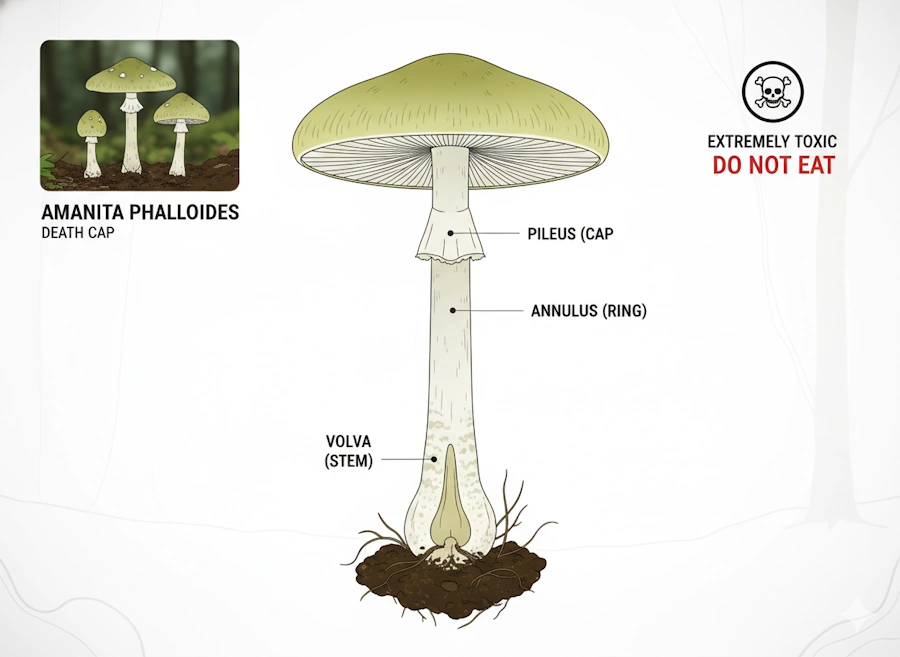
Understanding the death cap’s physical structure is essential for recognition, though this knowledge comes with a crucial caveat: the mushroom’s appearance is deceptively variable, and color alone cannot reliably identify it.
The cap
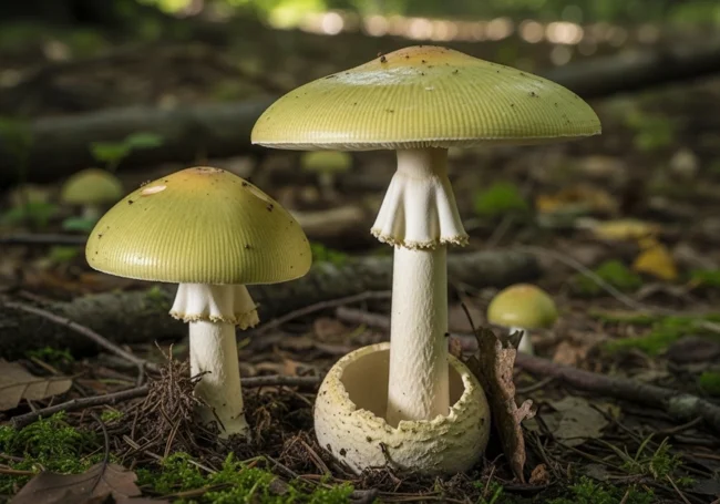
The pileus, or cap, typically measures between 5 and 15 centimeters across, though occasionally larger specimens appear. Young mushrooms begin with a rounded or hemispherical cap that gradually flattens to convex and eventually nearly planar as the fungus matures. The flesh inside is consistently white and does not discolor when cut.
The cap color is the aspect most commonly cited in popular descriptions, yet this is precisely where danger lies. Most commonly, the cap displays a pale yellowish-green or olive hue, sometimes with a metallic sheen that intensifies with age or drying. However, a pure white form exists and appears sporadically among green specimens. Intermediate colors occur regularly: tan, light brown, and grayish-brown caps have all been documented. Geographic location influences color intensity—populations in different regions display characteristic color ranges. This variability means that relying on cap color for identification is unreliable and potentially fatal.
Young death caps frequently bear patches of white tissue adhering to the cap surface. These are remnants of the universal veil, a membranous sheath that originally enveloped the entire young mushroom. In dry weather, these patches are particularly prominent, appearing as large creamy-white areas rather than the regularly scattered spots characteristic of other toxic Amanitas. However, rain and age can wash or wear away these distinctive markings.
The veil and volva
The universal veil is a crucial identifying feature of young Amanita specimens. When the mushroom first forms, it is completely enclosed within this membranous tissue, creating a ball-like structure that superficially resembles a puffball.
As the mushroom grows, pressure from the expanding cap causes the veil to tear. This rupture leaves two permanent structures: an annulus (partial veil) that sometimes remains as a delicate ring around the stem near the cap, and critically, a volva—a cup-like or bulbous structure that encircles the base of the stem where the mushroom appears from the ground.
The volva is the most chemically toxic part of the entire mushroom, containing extraordinarily concentrated levels of phallotoxins. In young or very small specimens, the volva may remain entirely buried beneath the soil surface, rendering it invisible to a forager who simply pulls up a mushroom. Complete excavation and careful observation of the base, revealing this distinctive cup structure, is absolutely necessary for proper identification.
Many tragic poisonings have resulted from foragers failing to unearth the mushroom completely and thus missing this diagnostic feature.
The stem and gills
The stipe, or stem, is typically white or pale cream-colored, occasionally with faint greenish tones. The diameter tapers from a slightly thicker base upward, creating a somewhat delicate appearance. The stem is generally hollow, particularly when the mushroom matures.
Beneath the cap, the gills extend from the center outward toward the cap margin in a characteristic radial pattern. The gills are free—meaning they do not attach to the stem but rather end before reaching it. They are consistently white, a feature shared with several edible species, which increases the risk of misidentification. When treated with concentrated sulfuric acid in laboratory conditions, the gills take on a pallid lilac or pink coloration, though this test is impractical for field identification.
Spore print
The spore print—the pattern created when gills deposit spores onto paper left overnight beneath the cap—is white, another feature common to many Amanita species and therefore not particularly diagnostic by itself. The individual spores are globular to egg-shaped, measuring 8-10 micrometers in length. Under a microscope, they stain blue with iodine, but again, this observation requires laboratory access.
Smell and taste
Young death caps at the button stage are essentially odorless or possess only a faint, barely perceptible honey-sweet smell. As the mushroom matures, this scent intensifies dramatically to become overwhelmingly pungent and unpleasant—a sickly-sweet, objectionable odor. However, many early-stage mushrooms retain their mild or absent odor, providing no olfactory warning.
Accounts from poisoning survivors indicate that the raw or cooked flesh tastes quite pleasant—a bitter-sweet, agreeable flavor that provides no hint of the toxins within.
This palatability arguably increases the danger: the mushroom offers no sensory warning that something is amiss.
Distinguishing from edible lookalikes
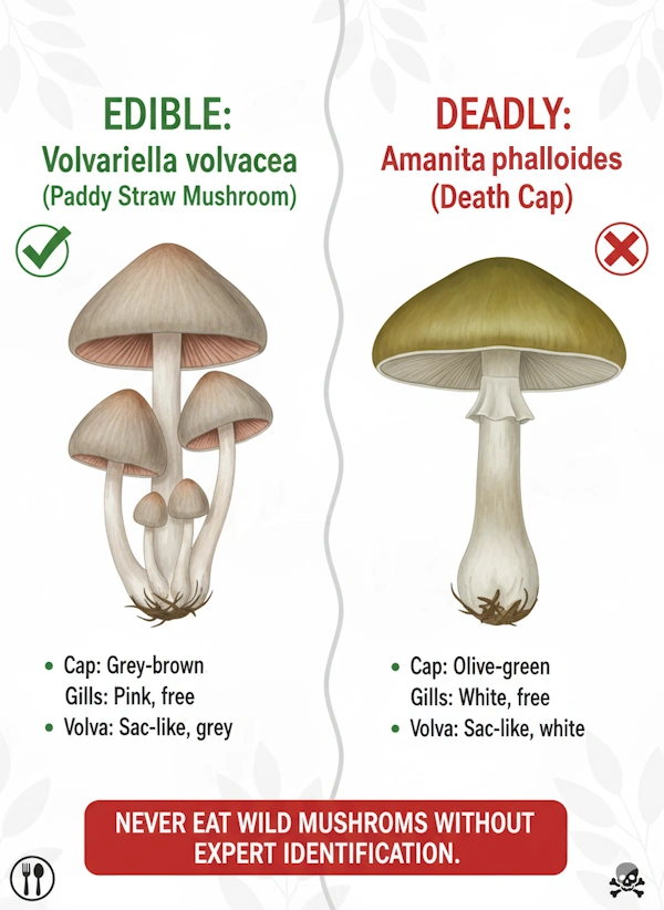
The death cap’s most dangerous mimics are edible species. The paddy straw mushroom (Volvariella volvacea) closely resembles the death cap in cap color and size and possesses a white cup-like structure at its base. However, the paddy straw has pink or salmon-colored spores rather than white, and the gills are pink rather than white.
Tragically, this distinction means little to individuals from Southeast Asia where paddy straw mushrooms are cultivated and consumed regularly but death caps do not occur naturally—when these foragers encounter death caps in North America or Europe, they instantly recognize what they believe to be the familiar mushroom they have eaten safely their entire lives.
Young death caps in the egg or button stage can be confused with edible puffballs. The critical test: slice the specimen lengthwise from top to bottom. An edible puffball presents a completely uniform, white interior throughout. A death cap “egg” reveals internal structure—most tellingly, the developing gills of the mature mushroom can be visible as half-moon-shaped formations, and the characteristic cup of the volva is apparent at the base.
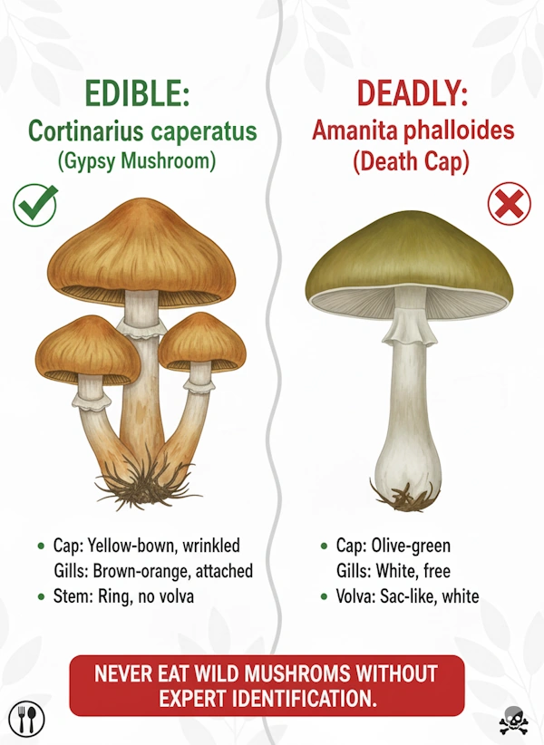
Mature brownish death caps have been mistaken for the gypsy mushroom (Cortinarius caperatus), which differs in possessing brown rather than white gills, a wrinkled rather than smooth cap surface, and brown spores instead of white.
The destroying angels (Amanita virosa, A. bisporigera, A. ocreata) and fool’s mushroom (A. verna) are closely related to the death cap and equally deadly, though typically lacking the green or yellow pigments characteristic of most death caps.
The toxins of death cap
The death cap is not merely toxic—it is a pharmaceutical factory of multiple poisons, each with distinct mechanisms of harm.
three classes of toxins
Researchers have identified three major groups of toxins in Amanita phalloides: amatoxins, phallotoxins, and virotoxins. However, their relative contributions to overall toxicity are markedly different.
Amatoxins are the primary killers. These heat-stable cyclic peptides consist of arrangements of eight amino acid residues organized in a specific three-dimensional structure. At least nine different amatoxins have been identified, with alpha-amanitin (α-amanitin) being overwhelmingly the most toxic to humans. The molecule’s three-dimensional shape is remarkably stable: it resists the high temperatures of cooking, freezing during storage, and the harsh acidic environment of the stomach. A single death cap mushroom can contain up to 15 milligrams of amatoxins, yet as little as 0.1-0.4 milligrams per kilogram of body weight can prove lethal. This means that consuming just half of a medium-sized mushroom contains sufficient amatoxin to kill an average adult.
Phallotoxins represent a secondary toxin class consisting of at least seven compounds, all containing seven similar peptide rings with particularly toxic bonds involving sulfur atoms and indole rings. Remarkably, despite being highly poisonous to liver cells, phallotoxins contribute little to death cap toxicity because they are not significantly absorbed through the intestinal wall and therefore do not reach the liver in sufficient quantity. The volva, however, contains extraordinarily concentrated phallotoxin levels.
Virotoxins represent a third identified toxin class whose precise role in human poisoning remains incompletely understood. Their contribution to overall toxicity appears minimal compared to amatoxins.
How alpha-amanitin destroys cells
The mechanism of action of alpha-amanitin is elegantly specific and catastrophically effective. At the molecular level, this toxin functions as an inhibitor of RNA polymerase II, an essential enzyme complex responsible for transcribing DNA into messenger RNA (mRNA). This enzyme operates by moving along the DNA strand, reading genetic instructions and synthesizing mRNA molecules that serve as templates for protein synthesis. Alpha-amanitin binds specifically to a highly conserved region called the bridge helix within the RNA polymerase II complex. This binding locks the bridge helix in place, dramatically slowing the enzyme’s movement along DNA from a normal rate of several thousand nucleotides per minute down to just a few nucleotides per minute.
The consequence is catastrophic: mRNA synthesis nearly stops. Without new mRNA, cells cannot synthesize the proteins necessary for maintaining basic cellular functions. Enzyme systems fail, structural proteins break down without replacement, and energy production halts. The cell enters a state of metabolic arrest and dies.
The mutation that makes Amanita phalloides immune to its own toxin is fascinating from an evolutionary perspective: the fungus’s own RNA polymerase II contains amino acid substitutions in the bridge helix region that prevent amatoxin binding while maintaining full enzymatic function. The mushroom manufactures a lethal poison while being completely insensitive to it—a biochemical loophole that allows the fungus to produce and accumulate toxins without self-harm.
Toxin distribution and concentration
The concentration of toxins varies among individual mushrooms collected from the same population, with well-developed fruiting bodies accumulating higher toxin levels than young specimens. The volva represents the most concentrated toxic zone, making contact with the base of the mushroom particularly dangerous. Young eggs of death caps, less developed in toxic chemistry, still contain lethal amounts of poison—there is no truly “safe” developmental stage. Toxin content varies geographically between populations as well, making it impossible to predict lethality based on appearance alone.
Critically, toxin concentrations do not diminish through cooking, freezing, pickling, drying, or any other food preparation method. Heat-stability makes these toxins particularly dangerous: traditional preservation and cooking methods that might render other mushroom toxins inactive have no effect on amatoxins.
Poisoning: the progression of a silent killer
Death cap poisoning is uniquely sinister because it creates a false sense of security. The toxins are not irritating to the gastrointestinal tract in the immediate aftermath of ingestion, leading to a prolonged asymptomatic period during which massive organ damage is silently occurring.
Phase one: the latent period (6-40 hours, average 10 hours)
After ingestion, hours pass with no symptoms whatsoever. The victim feels completely normal, experiences no nausea, no pain, no warning whatsoever. This is the phase where most people do not seek medical attention because they have no reason to suspect anything is wrong.
During this deceptively peaceful period, amatoxins are being rapidly absorbed through the intestinal wall into the bloodstream. The portal blood system carries the toxins directly to the liver, where they accumulate in hepatocytes. At the molecular level, RNA polymerase II is already bound by amatoxin, mRNA synthesis is collapsing, and hepatocytes are beginning to die. Kidney cells are accumulating toxins as well.
The duration of this latent phase is variable and unpredictable. Some individuals manifest symptoms after 6 hours; others remain asymptomatic for up to 40 hours. This variability reflects differences in absorption rates, individual metabolism, and toxin concentration in the specific mushroom consumed. The prolonged latency period is diagnostically treacherous: by the time symptoms appear, gastric lavage and activated charcoal are largely ineffective because the stomach has already emptied and the toxins have been absorbed.
Phase two: gastrointestinal illness (12-24 hours after ingestion)
Suddenly, the asymptomatic phase breaks. Violently. Victims experience acute onset of severe nausea, intense abdominal cramping, and profuse watery diarrhea. Vomiting begins, and in severe cases, the diarrhea and vomit become grossly bloody. The gastrointestinal symptoms are caused by phallotoxins damaging the cellular membranes of enterocytes—the epithelial cells lining the intestines.
This phase can produce severe dehydration, electrolyte abnormalities, acid-base disturbances, dangerously low blood glucose, and hypotension. Victims often present to emergency departments during this phase, and here begins another deadly deception: the gastrointestinal symptoms resemble common viral gastroenteritis, food poisoning, or cholera. Many emergency physicians lack awareness that this particular cluster of gastrointestinal symptoms arising 12 or more hours after mushroom ingestion is characteristic of amatoxin poisoning, not bacterial contamination or viral illness.
The second phase typically lasts 12 to 24 hours. As intestinal contents are purged, gastrointestinal symptoms gradually diminish. The vomiting stops, the diarrhea ceases, the abdominal pain subsides. The victim and their physicians experience a profound sense of relief. Many patients are discharged from medical care at this point because they appear to be recovering. This apparent recovery is devastatingly false.
Phase three: the silent progression (24-72 hours after ingestion)
During this interval, the patient feels surprisingly well—better than during the gastrointestinal phase. There is no pain, no visible symptoms of severe illness. However, this is the phase during which hepatic and renal failure is rapidly progressing. Amatoxins have been accumulating in the liver and kidneys for days, and the toxin load has reached critical levels. Hepatocytes are dying en masse through necrosis. The liver swells with inflammation and cellular death. Kidney tubules are damaged, and filtration capacity declines.
Liver enzyme levels (AST, ALT, bilirubin) begin to rise dramatically. Blood tests show evidence of reduced protein synthesis. Prothrombin time extends as the liver loses capacity to produce clotting factors. These changes are evident on blood tests but produce no symptoms that the patient can feel. The patient may feel progressively fatigued, but this is often attributed to the stress of recent illness rather than recognized as a sign of incipient organ failure.
Phase four: hepatorenal failure and death (72+ hours after ingestion)
Three to seven days after ingestion, the catastrophic failure phase begins. The liver damage becomes manifest clinically: jaundice develops as bilirubin accumulates (yellowing of skin and sclera of the eyes), coagulopathy emerges (blood becomes unable to clot properly), hepatic encephalopathy develops as ammonia and other neurotoxic metabolites accumulate due to failed liver detoxification.
Patients develop altered mental status, confusion, delirium. Seizures may occur. The person may fall into a coma. Kidney function collapses simultaneously—urine output drops (oliguria), and toxins accumulate in the bloodstream. Hemorrhage occurs as coagulation failure allows bleeding into tissues. Cardiovascular instability develops. Multi-organ failure becomes apparent.
Without liver transplantation, death occurs within 5 to 8 days of ingestion. Post-mortem examination reveals a massively enlarged, red, edematous liver, often with hemorrhage, and severe renal damage.
The mortality rate after death cap poisoning ranges from 10 to 20 percent even with modern medical intervention, and this improvement from historical rates of 60-70 percent reflects advances in intensive care, dialysis, antibiotic therapy, and liver transplantation. Notably, approximately 20-79 percent of patients who survive amatoxin poisoning develop chronic liver disease, meaning recovery is rarely complete even when death is avoided.
Critical factors determining outcomes
The dose consumed is the most important determinant of outcome. As little as 30 grams (roughly half a medium mushroom cap) can be lethal. Consuming an entire mushroom—50 grams—almost certainly will be fatal without immediate liver transplantation.
Individual variation in susceptibility is significant. Children appear to absorb higher proportions of toxin relative to their body weight and have substantially higher morbidity and mortality than adults. Pre-existing liver disease dramatically worsens prognosis. General health status and immune function influence outcomes.
The time interval between ingestion and initiation of aggressive medical treatment is crucial. Patients treated within 24 hours have better outcomes than those treated later, though even this window is often missed because symptoms are delayed.
Why death cap poisoning is so dangerous
Multiple factors converge to make amatoxin poisoning a uniquely lethal threat:
- First, the prolonged asymptomatic phase creates a false sense of safety and delays medical intervention until irreversible damage has occurred.
- Second, the delayed gastrointestinal symptoms are nonspecific and mimmic common illnesses, leading to misdiagnosis or missed diagnosis.
- Third, the apparent recovery during phase three is a deadly trap that leads to premature discharge from medical care just before critical failure begins.
- Fourth, amatoxins are completely resistant to all food preparation methods—no amount of cooking, boiling, drying, freezing, or fermenting reduces their toxicity.
- Fifth, the mushroom’s appearance is deceivingly variable and closely resembles edible species commonly consumed in the regions to which it has invaded.
- Sixth, many people remain unaware of the mushroom’s existence in their region and thus include no death cap recognition in their foraging knowledge.

