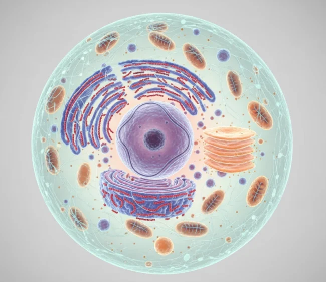
The cell is the basic unit of all living organisms. It is a complex system in which each part performs specific functions. Just as a city consists of various buildings and institutions, a cell contains numerous organelles, each responsible for particular life processes.
Studying cell structure: how it all started
“Nature has created all beings according to a single structural plan, identical in principle but infinitely varied in detail.” This was the opinion expressed in the early 19th century by the French zoologist Étienne Geoffroy Saint-Hilaire.
Persistently, despite widespread prejudice, Saint-Hilaire defended his scientific views. In a famous public debate with Georges Cuvier, Saint-Hilaire attempted to prove the similarity between the structural plans of vertebrates and cephalopod mollusks.
Cuvier brilliantly and convincingly refuted Saint-Hilaire’s arguments. And yet, today we can say that Saint-Hilaire was… right. He was just looking for a unified structural plan at the level of entire organisms—ones that were too distant from each other. However, at the cellular level, the structural unity of all living beings is universally recognized.
Cells of all living organisms are similar (homologous) to each other. This is also one of the principles of cell theory. Just as an organism consists of individual organs, a cell consists of many parts responsible for nutrition, excretion, reproduction, etc. These structural components of the cell are called organelles. In plant, animal, and fungal cells, we find the same organelles (though there are differences).
Scientists have been discovering and studying new organelles for decades and even centuries. The study of cells can be divided into two eras: light microscopy and electron microscopy.
The naked eye can distinguish objects no smaller than one-tenth of a millimeter. The microscope, invented in 1590 by the Dutch mechanics Hans and Zacharias Janssen, made it possible to extend this visibility limit by tens and later hundreds of times.
Today, the best light microscopes allow us to see details 500 times smaller than what the naked eye can detect—but no smaller! Light waves simply bend around such “smallness,” just as ocean waves roll over medium-sized boulders without “noticing” them. The only way to resolve finer details is to use shorter waves that can penetrate smaller “gaps” and interact with even the tiniest “pebbles.”
The electron microscope, developed in the 1930s, allows us to see far more cellular details than a light microscope. Instead of visible light, the object is illuminated by a focused beam of electrons. Modern electron microscopes have extended the limit of visibility another 2,000 times beyond that of light microscopes. This resolution is now approximately equal to the diameter of a hydrogen atom.
Cell nucleus
The nucleus can be called the “command center” of the cell. It is the most noticeable structure, first observed by early cell researchers in the 17th century. Just as a central office stores all important company documents, the nucleus contains the cell’s genetic material—DNA, which holds the organism’s hereditary information.
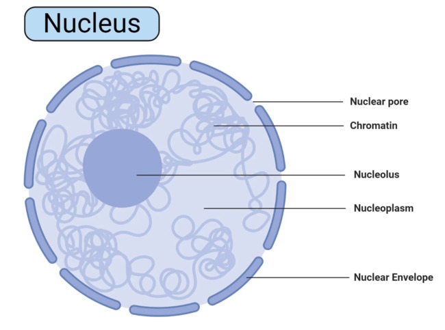
The nucleus is surrounded by a double membrane—the nuclear envelope—which has numerous pores. These pores function as “checkpoint stations,” allowing the nucleus to send its “instructions” in the form of RNA molecules into the cytoplasm and regulate the entire cell’s activity. One could say these pores work like post offices, facilitating the two-way exchange of molecules between the nucleus and the cytoplasm.
Inside the nucleus is a specialized structure where ribosomes, the cell’s “protein factories,” are assembled. The nucleus also contains chromatin, the substance that forms chromosomes during cell division. Chromatin can be compared to a tangled ball of threads that straightens out and becomes visible as distinct chromosomes when the cell divides.
In higher organisms, each cell usually has one nucleus, although multinucleated cells exist (for example, liver cells). Some cells, such as human red blood cells, lose their nucleus during development. Without a nucleus, a cell cannot divide and has a limited lifespan.
Cell membranes
Biologists have long suspected that every cell is surrounded by a thin “skin,” a membrane that separates it from the external environment. However, it was only in the 1950s, with the help of an electron microscope, that this membrane could be observed. So, what exactly is this cellular “skin”?
To get a clear idea of it, let’s recall an ordinary soap bubble. Water gradually flows down, thinning the bubble’s wall. Then, rainbow streaks start running down from the top. This means that the thickness of the soap film has reached just a few hundred molecules and has become comparable to the wavelength of light.
As the film gets even thinner, the entire spectrum of colors flashes across the bubble several times.
Then, something remarkable happens. A “hole” appears at the top of the bubble, rapidly expanding. The bubble bursts. If the bubble is suspended in the air, at some point, it may seem that only the lower hemisphere remains. However, the upper part of the soap film does not actually disappear. It simply becomes so thin—just two or three molecules thick—that light waves pass through it as if there were no barrier at all!
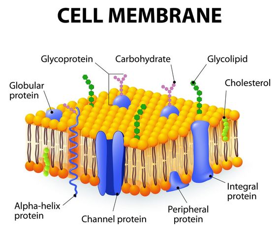
Each living cell is wrapped in an equally thin film (a membrane) just two molecules thick. In terms of viscosity, the membrane is similar to olive oil. Protein molecules are embedded in this lipid film. These proteins are not fixed in place but float freely within the membrane. They serve as the membrane’s “checkpoint controllers,” its “gatekeepers.” Proteins not only allow “invited guests” into the cell but also expel “unwanted visitors.” Some do this skillfully. The membrane also has “doorbells” (which are also proteins) that transmit signals inside the cell. These proteins function like “apartment numbers,” enabling cells to recognize each other.
In animal cells, an additional “coating” of carbohydrates lies on top of the outer cell membrane, giving it extra strength. In plant cells, in addition to the membrane, there is also a thick cell wall made of cellulose.
The membrane not only encases the cell but also partitions it (biologists say it “compartmentalizes” the cell) into separate sections, each carrying out its own chemical processes. Within these compartments, the cell produces its proteins, fats, and carbohydrates. This internal cellular labyrinth of membranes, with its “tunnels,” canals, and cavities, was discovered in 1945. It was named the endoplasmic reticulum. In a way, the cell is divided into “kitchens,” “offices,” “dining rooms,” and so on.
Imagine if people stopped building interior walls in houses—living in such spaces would be far less convenient! Similarly, when looking at cells, it is worth noting that cells of nucleus-free organisms—bacteria and cyanobacteria—are structured with minimal internal compartments.
Lysosomes
Lysosomes were discovered in 1955. These are tiny membrane-bound vesicles filled with special protein enzymes.
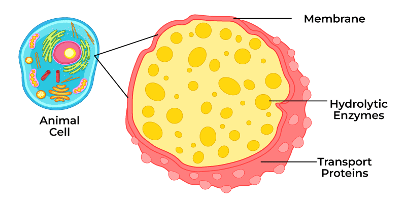
These proteins are so effective at breaking down and digesting organic substances that if they were “released” from the lysosomes, the cell would “digest itself.” Lysosomes function like intracellular “stomachs”. They digest not only food entering the cell but also damaged parts of the cell itself. Lysosomes also have other functions.
For example, the male reproductive cell uses lysosomes to “burn” a path through the egg cell’s membrane for fertilization. During a tadpole’s transformation into an adult frog, lysosomes “devour” its tail.
Golgi Complex
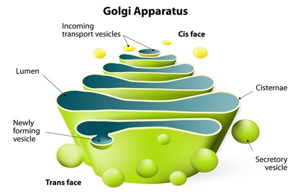
It is also called the Golgi apparatus, named after the Italian scientist who discovered it in 1898. This organelle collects and “packages” substances produced by the cell (proteins, fats, carbohydrates), usually for export to different organs. Lysosomes are also produced here. The Golgi complex consists of flat membrane-bound vesicles stacked on top of each other like pancakes.
Mitochondria
Billions of years ago, bacteria-like organisms found an unusual habitat—they settled inside the cells of other living organisms. Over time, the “hosts” and their “tenants” adapted to each other, and eventually, they became so interdependent that they could no longer survive without one another. This form of mutual assistance in nature is known as symbiosis.
This partnership proved so beneficial that today, these “lodgers” continue to live in nearly all cells of plants, fungi, and animals—including our own cells—becoming an essential part of them. They are called mitochondria, and in plants, plastids as well.
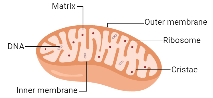
From their former independence, mitochondria retain only relative autonomy. They have their own genetic information stored in DNA and can synthesize some of their own proteins. However, this is not enough for them to reproduce freely outside the cell. New mitochondria (and plastids, which will be discussed later) arise by dividing existing ones.
Mitochondria are known as the “batteries of life,” the “power plants of the cell.” Cellular respiration occurs in mitochondria. Without fire and smoke, they efficiently “burn” nutrients and convert the released energy into ATP, which is then distributed in a convenient “packaged” form for all cellular needs. The efficiency of mitochondria is remarkably high—about 50%, while internal combustion engines operate at around 33%.
A cell can contain anywhere from one to several thousand mitochondria—the more work a cell must perform, the more space mitochondria occupy (up to 40% of the total cell volume).
Vacuole
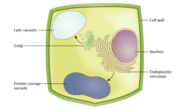
The vacuole in plant cells serves as a storage for cell sap. A vacuole for a cell is like a pantry for a thrifty owner. However, this “pantry” can grow to enormous sizes, sometimes occupying up to 90% of a plant cell’s volume. What do cells store in their “storage rooms”? Usually, salts, vitamins, and sugars, and sometimes soluble proteins. A cell can also deposit toxic metabolic byproducts in the vacuole, such as nicotine. Large vacuoles are not found in animal cells.
Plastids
Plastids share a similar history with mitochondria: from accidental “tenants,” they became an integral part of the cell. The internal structure of mitochondria and plastids is similar. However, while mitochondria are believed to have evolved from bacteria, plastids are thought to have originated from blue-green algae. Plastids are found only in plant cells.
When we see autumn forests changing from green to yellow and red or when a potato tuber turns green after exposure to light, we are witnessing the transformation of one type of plastid into another.
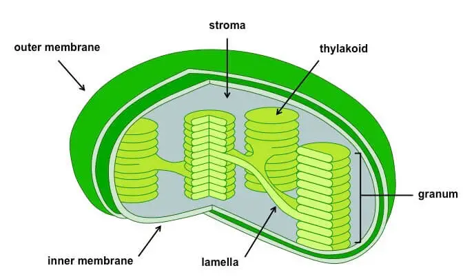
Green plastids are called chloroplasts. Photosynthesis occurs within them (see the article “Photosynthesis”). They function like tiny solar panels. A plant cell can contain anywhere from one to several hundred chloroplasts.
Chloroplasts can move independently within the cell. For example, to avoid excessive light exposure, they hide behind the “curtains” of cell membranes. The chloroplast in the green alga Mougeotia behaves in an interesting way: under dim light, it exposes its broad surface to capture more light, while under intense light, it “hides” by turning sideways.
Colorless plastids that store nutrients such as oils and starch are called leucoplasts. The leucoplasts in potato tubers, filled with starch, transform into chloroplasts when exposed to light, causing the tuber to turn green.
When a tree prepares for leaf fall, its chloroplasts turn into brightly colored chromoplasts. The same transformation happens when fruits ripen, turning from green to their mature color. Chromoplasts also give flowers their vibrant petal colors.
Can a cell be seen?
The body of an adult human consists of approximately 100 trillion cells. But how many cells do you think a newly fertilized chicken egg contains? It turns out that, like a bird’s egg, any egg cell is a single cell surrounded by a dense membrane. In many cases, distinguishing egg cells is not difficult.
In a spring pond, it is easy to find transparent “chains” of frog spawn, with numerous black dots inside. Each of these black dots in the light-yellow egg mass is also a single cell—an egg cell.
Organisms consisting of a single cell (such as amoebas, ciliates, and many algae) are often visible to the naked eye. Their length can reach several millimeters, or sometimes even centimeters.
In the pulp of fruits like oranges, if you look closely, you can distinguish individual large cells. In the central part of the fruit, the visible juicy cells can reach up to 1 cm in diameter. The cells of multicellular animals are among the smallest, but in some cases, they can be seen. For example, if you take a drop of blood from an axolotl (a type of amphibian that some people keep in home aquariums) and release it into a small amount of water on a glass surface, you will see tiny oval clumps—blood cells—against a dark background.
Cells in a test tube in a laboratory
In 1907, the American biologist Ross Harrison reported an astonishing fact: he managed to keep frog embryo cells alive in a test tube for several weeks. This marked the beginning of growing cells outside the body.
And in 1950, a human cell culture was obtained for the first time in the United States. Cancerous tumor cells were taken from an African American woman, Henrietta Lacks (although in many textbooks, this woman is mistakenly referred to as Helen Lane). The cell culture was named after her initials, HeLa. Over time, these cells became the “standard” against which biologists worldwide compare their experimental results. Interestingly, after years of being cultured, HeLa cells have developed a high level of survivability. If they accidentally enter another cell culture, these aggressive invaders quickly displace the original “residents” and take over.
HeLa cells continue to live in laboratories, even though their original host passed away many decades ago.
Leave a Reply