General Virology: What Are Viruses, Are They Alive Or Not?
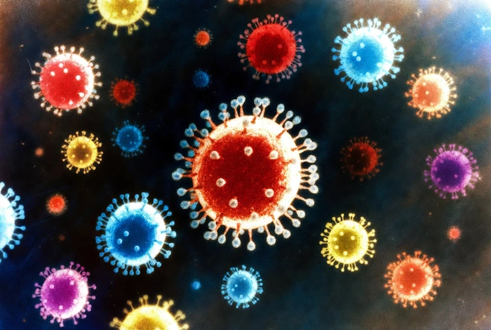
We often think of predators as big animals—lions, sharks, or bears. But the most efficient killers on Earth are invisible. They are tiny enough that countless individual units could occupy the tip of a single pin. They are Viruses.
But here is the mystery: Are they even alive? They don’t breathe. They don’t eat. They can crystallize like salt. Yet, once they enter your body, they hijack your cells and turn them into factories to build more of themselves.
In this lesson, we will explore the alien architecture of viruses, how they hack the genetic code, and the immune war your body fights every day to stop them.
[INTERACTIVE TOOL: VIRUS BUILDER]
Virus Builder Lab
1. Genetic Code
2. Capsid Shape (Protein)
3. Armor (Envelope)
Experiment: Build your own virus. Choose a DNA or RNA core. Pick a Capsid shape (Icosahedral vs. Helical). Add an Envelope. See if it can infect a human cell.
What Are Viruses?
Viruses are microscopic infectious agents that occupy a unique place in biology. They are not considered living organisms in the traditional sense because they lack cellular structure and cannot reproduce independently. Instead, viruses are obligate intracellular parasites, meaning they must infect a living cell—such as a bacterium, plant, animal, or human—to replicate.
1. Basic Structure of Viruses

A complete virus particle outside a host cell is called a virion. Virions consist of two essential components:
- Genetic Material: Viruses contain either DNA (deoxyribonucleic acid) or RNA (ribonucleic acid), but never both. This genetic material contains the code that enables the formation of additional viruses.
- Protein Coat (Capsid): The genetic material is enclosed in a protective shell made of proteins called a capsid. The capsid is composed of individual protein subunits called capsomeres, which assemble into precise geometric shapes, such as helices or polyhedral forms.
Some viruses also have an outer lipid envelope, derived from the host cell membrane, which helps them evade the host’s immune system and enter new cells more efficiently.
2. How Viruses Reproduce
Viruses cannot reproduce on their own. Instead, they hijack the machinery of a host cell to make copies of themselves. The process typically involves the following steps:
- Attachment: The virus binds to specific receptors on the surface of a host cell.
- Entry: The virus enters the cell, either by fusing with the cell membrane or being engulfed by the cell.
- Replication: The viral genetic material takes over the host cell’s machinery, directing it to produce viral components (nucleic acids and proteins).
- Assembly: Newly created virus particles are put together within the infected host cell.
- Release: The newly formed viruses exit the host cell, often destroying it in the process, and go on to infect other cells.
This cycle allows viruses to spread rapidly within an organism.
3. Why Are Viruses Not Considered Alive?
Viruses are often described as being “on the edge of life” because they exhibit some characteristics of living organisms but lack others:
- They cannot reproduce independently—they require a host cell.
- They do not metabolize or grow—they do not carry out chemical reactions to obtain energy.
- They do not respond to stimuli—they lack the structures needed for sensory perception.
However, they do evolve through mutations and natural selection, much like living organisms.
4. The Role of Viruses in Nature and Medicine
Viruses play a crucial role in ecosystems:
- They control populations of bacteria and other microorganisms.
- They drive evolution by transferring genes between species (horizontal gene transfer).
- Some viruses are used in gene therapy and vaccine development.
However, viruses are also responsible for many diseases, including the common cold, influenza, HIV/AIDS, COVID-19, and Ebola.
5. Classification of Viruses (The Technical Details)
Viruses are classified based on:
- Type of genetic material (DNA or RNA, single-stranded or double-stranded).
- Shape and structure (helical, icosahedral, or complex).
- Presence or absence of an envelope.
- Host range (which organisms they infect).
Distinctive features:
- They contain only one type of nucleic acid (RNA or DNA).
- They lack their own protein-synthesizing and energy systems (No Ribosomes, No Mitochondria).
- They lack cellular organization.
- They reproduce through a disjunctive method: Protein parts and DNA parts are made in different places and assembled later (like a car factory).
- Obligate parasitism occurs at the genetic level.
- Viruses pass through bacterial filters (they are much smaller than bacteria).
Shapes and Sizes: Virions can be Round, Rod-shaped, Polygonal, or Filamentous. Their sizes range from 15 nm (tiny) to 400 nm (large).
The Invasion (How Virus Interacts with the Host Cell)
When a virus meets a cell, it isn’t a random event: it involves a highly specific “lock‑and‑key” type interaction. The virus has proteins (Keys) that fit perfectly into receptors (Locks) on your cell surface.
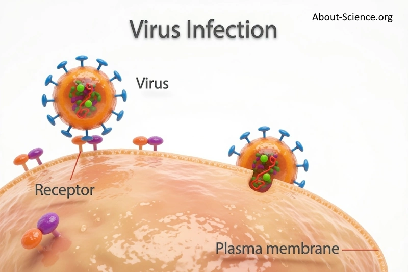
Look at the picture. The Main Players:
1. On the left side, you see a round, spiky orange ball labeled “Virus.” That’s our villain—or, scientifically speaking, the virion, which is the complete virus particle outside a cell. It’s got a protein coat (called a capsid) with those blue spikes sticking out, which are like keys. Inside, there’s genetic material (the green squiggly stuff), which could be DNA or RNA, depending on the virus.
2. The Target: Below the virus is a curved, pinkish surface labeled “Plasma membrane.” This is the outer boundary of a host cell, like the skin of a balloon. It’s made of lipids and proteins, and it’s the cell’s first line of defense. Sticking out from it are little purple and orange knobs—one is specifically labeled “Receptor.” Receptors are proteins on the cell surface that normally help with communication or nutrient uptake, but viruses exploit them.
3. The Attachment Phase: Look closely: The virus on the left is hovering near the membrane, with an arrow pointing to the receptor. This shows the virus approaching and binding to the receptor using its spikes (often called glycoproteins or attachment proteins). It’s like a lock and key—the spike fits perfectly into the receptor. This is the first step of infection: attachment. Without this match, the virus can’t get in.
4. The Entry Phase: Now, shift your eyes to the right side. There’s another virus that’s already attached and starting to sink into the membrane. This illustrates endocytosis or fusion, where the virus either gets swallowed by the membrane (forming a bubble inside the cell) or fuses its envelope with the membrane to release its genetic material directly. Once inside, the virus’s genome takes over the cell’s machinery to replicate, assemble new viruses, and eventually burst out to infect more cells.
Why does this matter? This process is how viruses like influenza, HIV, or even coronaviruses cause diseases. It helps scientists design vaccines (which train your immune system to block those spikes) or antiviral drugs (that jam the lock).
The 4 Types of Virus Infection
- Productive Infection: The virus multiplies, the cell dies. (e.g., Flu).
- Abortive Infection: The virus tries to enter, but fails. The cell survives.
- Latent Infection: The virus enters and hides. It sleeps in your DNA without killing the cell (e.g., Herpes).
- Transformation: The virus changes the cell’s DNA, potentially causing it to become cancerous (e.g., HPV).
The Virus Lifecycle Steps (Deep Dive)
- Adsorption: The virus sticks to the cell.
- Penetration (Viropexis): The cell swallows the virus (Endocytosis).
- Deproteinization (“Undressing”): The cell digests the virus’s protein coat, releasing the naked Viral DNA/RNA into the cytoplasm.
- Replication: The viral DNA travels to the nucleus. It forces the cell to copy it.
- Assembly: The cell builds new Capsids and stuffs the new DNA inside them.
- Release:
- Lytic Release: The cell explodes (Lysis), releasing thousands of viruses.
- Budding: The virus pushes through the membrane, stealing a piece of “skin” (Envelope) as it leaves. This keeps the cell alive longer to make more viruses.
How We Study Viruses (Cultivation)
You cannot grow viruses on a Petri dish like bacteria (because they need living cells to eat). So, how do scientists study them?
1. Laboratory Animals: Injecting mice or rabbits. (Old school method).
2. Chicken Embryos: Injecting a fertilized egg. The virus grows inside the developing chick. (This is how we make Flu Vaccines!).
3. Tissue Culture (The Modern Way): Scientists grow sheets of human or animal cells in plastic flasks. They infect these cells and watch them die under a microscope.
- The Cytopathic Effect (CPE): We know the virus is working if the cells round up, detach, fuse together (Syncytia), or explode.
The War Inside (Antiviral Immunity)
Your body doesn’t give up without a fight. You have a complex defense system.
1. The First Responder: Interferon. When a cell is infected, it screams for help by releasing a chemical called Interferon. It is a type of a human protein and it is designed to fight viral infections.
- What it does: It warns neighboring cells: “I’m infected! Shut down your factories!”
- This stops the virus from spreading to healthy cells.
2. The Sniper: Antibodies (Humoral Immunity). Your B-Cells shoot Y-shaped proteins called Antibodies.
- Neutralization: They stick to the virus’s “Keys,” so it can’t unlock your cells.
- Opsonization: They act like a “Tag” that tells macrophages (eater cells) to come and swallow the virus.
3. The Assassin: Cytotoxic T-Cells (Cellular Immunity). Antibodies can’t reach a virus inside a cell. That is where T-Cells come in.
- The infected cell holds up a flag (Antigen Presentation) saying: “I have a virus inside me.”
- The Killer T-Cell sees the flag, docks with the cell, and injects toxins that cause the cell to self-destruct (Apoptosis). It sacrifices the one to save the many.
Immunity Memory
Why do you only get Chickenpox once? Your body keeps Memory Cells. If the same virus enters 10 years later, your body remembers the specific antibody shape and produces millions of them instantly, killing the virus before you even feel sick.
Summary of Key Terms
- Virion: A complete virus particle outside a cell.
- Capsid: The protein shell protecting the viral DNA/RNA.
- Obligate Parasite: An organism that cannot reproduce without a host.
- Interferon: A chemical alarm signal sent by infected cells.
- Lysis: The bursting/death of a cell.
🎓 Quiz: General Virology
1. Why are viruses NOT considered fully alive?
- A) They are too small
- B) They can kill humans
- C) They cannot reproduce without a host cell
- D) They are made of plastic
👉 Click to check answer
They lack the machinery (ribosomes) to multiply on their own.
2. What is the protein shell of a virus called?
- A) The Envelope
- B) The Capsid
- C) The Membrane
- D) The Wall
👉 Click to check answer
It protects the genetic material (DNA or RNA).
3. Which method is used to grow viruses in the lab?
- A) Agar Plates
- B) Sugar Water
- C) Living Tissue Cultures
- D) Soil samples
👉 Click to check answer
Since viruses need living cells to replicate, simple agar plates won’t work.
4. What does “Interferon” do?
- A) Kills the virus directly
- B) Warns neighboring cells to protect themselves
- C) Feeds the virus
- D) Builds new cells
👉 Click to check answer
It inhibits viral replication in nearby cells.
5. What is a “Latent Infection”?
- A) The virus kills the cell immediately
- B) The virus fails to enter
- C) The virus sleeps in the cell without killing it
- D) The virus turns into a bacteria
👉 Click to check answer
Like Herpes, it can reactivate years later.
Sources & References
- Knipe, D.M. and Howley, P.M. (eds) (2023) Fields Virology: Fundamentals. 7th edn. Philadelphia: Wolters Kluwer.
- Kumar, S. (2025) Textbook of General Virology. 1st edn. Boca Raton: CRC Press.
- Flint, S.J. et al. (2015) Principles of Virology: Molecular Biology, Pathogenesis, and Control. 4th edn. Washington, DC: ASM Press.
- Murray, P.R., Rosenthal, K.S. and Pfaller, M.A. (2020) Medical Microbiology. 9th edn. Philadelphia: Elsevier. (Chapters on general virology and viral pathogenesis).
- Netherton, C.L. and Wileman, T. (2007) ‘Virus factories, double membrane vesicles and viroplasm’, Journal of General Virology, 88(10), pp. 2367–2376.
- Routh, A. and Johnson, J.E. (2024) ‘Viral replication organelles: the highly complex and programmed replication machinery’, Frontiers in Microbiology, 15, p. 1450060.
- Ahlquist, P. (2006) ‘Parallels among positive-strand RNA viruses, reverse-transcribing viruses and double-stranded RNA viruses’, Nature Reviews Microbiology, 4(5), pp. 371–382.
- Forterre, P. (2016) ‘To be or not to be alive: How recent discoveries challenge the traditional definitions of viruses and life’, Studies in History and Philosophy of Biological and Biomedical Sciences, 59, pp. 100–108.
- Moreira, D. and López-García, P. (2009) ‘Ten reasons to exclude viruses from the tree of life’, Nature Reviews Microbiology, 7(4), pp. 306–311.
- Mahy, B.W.J. and Van Regenmortel, M.H.V. (eds) (2009) Desk Encyclopedia of General Virology. Amsterdam: Elsevier. (Sections on virus cultivation, embryonated eggs, and cell culture).
- Freshney, R.I. (2016) Culture of Animal Cells: A Manual of Basic Technique and Specialized Applications. 7th edn. Hoboken: Wiley-Blackwell. (Chapters on viral infection and cytopathic effects).
- Janeway, C.A. et al. (2017) Immunobiology: The Immune System in Health and Disease. 9th edn. New York: Garland Science. (Chapters on antiviral immunity, interferons, and cytotoxic T cells).
- Samuel, C.E. (2001) ‘Antiviral actions of interferons’, Clinical Microbiology Reviews, 14(4), pp. 778–809.
- Garcia-Sastre, A. and Biron, C.A. (2006) ‘Type 1 interferons and the virus–host relationship: a delicate balance’, Journal of Experimental Medicine, 203(5), pp. 1363–1371.
- Seiler, P. et al. (2006) ‘Sendai virus infection induces efficient adaptive immunity independent of type I interferons’, Journal of Virology, 80(9), pp. 4532–4542.
- Molinari, N.A. et al. (2009) ‘Dendritic cells in innate and adaptive immune responses against influenza virus’, Infection and Immunity, 77(9), pp. 3865–3874.
- Baltimore, D. (1971) ‘Expression of animal virus genomes’, Bacteriological Reviews, 35(3), pp. 235–241. (Foundational Baltimore classification).
- Venter, E., Delaney, A. and Fletcher, T. (2018) ‘Replication and expression strategies of viruses’, in Encyclopedia of Virology. 4th edn. London: Academic Press, pp. 382–393.